Notice: Function _load_textdomain_just_in_time was called incorrectly. Translation loading for the acf domain was triggered too early. This is usually an indicator for some code in the plugin or theme running too early. Translations should be loaded at the init action or later. Please see Debugging in WordPress for more information. (This message was added in version 6.7.0.) in /home/precisio/public_html/wp-includes/functions.php on line 6121
Endoscopic Endonasal Pituitary Surgery Singapore | Dr Teo Kejia

Endoscopic Endonasal Pituitary Surgery in Singapore

Living with a pituitary tumour can be challenging. Symptoms such as vision changes, hormonal imbalances, or headaches may occur when hormone production is affected. The thought of brain surgery often brings concerns about large incisions, hospital stays, and recovery. For many patients in Singapore, understanding available treatment options—including minimally invasive techniques—is an important part of managing these conditions.

Endoscopic endonasal pituitary surgery is a specialised surgical approach used to treat pituitary tumours and certain other skull base conditions. The procedure accesses the pituitary gland through the nasal passages, avoiding external incisions. This technique aims to reduce trauma to healthy brain tissue by working through natural passages rather than requiring large openings in the skull.

This guide provides an overview of endoscopic endonasal pituitary surgery to support informed decision-making about treatment options.

doctor img
Dr Teo Kejia (张哿佳医生)

MBBS (SG) MRCS (Edin) IFAANS FAMS (Neuro Surg) FRCS Ed (Neuro Surg)

pexels mart production 7089331

What is Endoscopic Endonasal Pituitary Surgery?

Endoscopic endonasal pituitary surgery, also known as transsphenoidal hypophysectomy, is a minimally invasive neurosurgical technique used to remove pituitary tumours and selected lesions at the skull base. The procedure is performed using an endoscope—a thin, rigid tube equipped with a high-definition camera and light source—which is inserted through the nostrils to reach the pituitary gland.

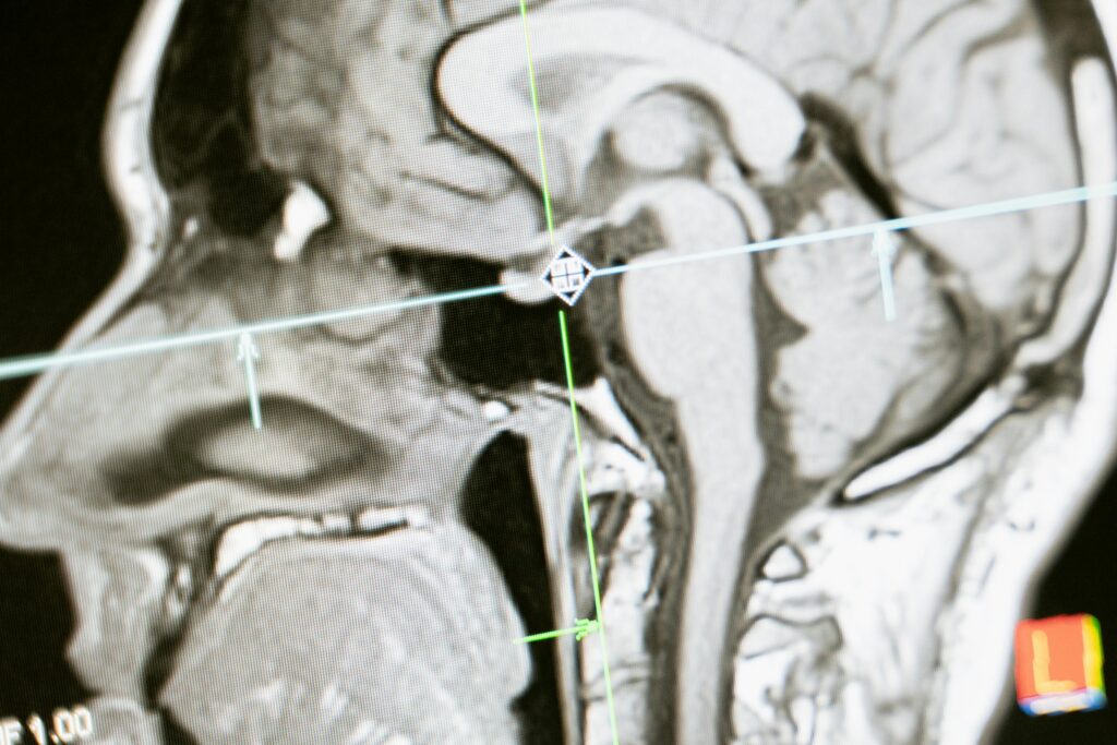
The pituitary gland is located within a small bony cavity known as the sella turcica, positioned behind the nose and between the eyes. Traditional surgical approaches to this region often required a craniotomy. In contrast, the endoscopic endonasal approach accesses the pituitary gland through the sphenoid sinus, a natural air-filled space located behind the nasal cavity.

Unlike traditional surgery, this approach provides a magnified, well-lit view of the surgical target, allowing the neurosurgeon to navigate with precision. It may be considered for the management of:

  • Pituitary adenomas, including functioning and non-functioning tumours
  • Craniopharyngiomas, which are rare tumours near the pituitary gland
  • Rathke’s cleft cysts (fluid-filled sacs in the pituitary region)
  • Brain cysts and Arachnoid cysts located in the sellar region
  • Selected meningiomas (slow-growing tumours forming on the protective layers of the brain)
  • Chordomas, which are rare bone tumours at the skull base
  • Cerebrospinal fluid (CSF) leaks, where protective fluid escapes from the skull base

Potential Candidates

You may be considered for this approach if you have:

  • Pituitary adenomas causing symptoms: Tumours compressing the optic nerves or producing excess hormones affecting growth and metabolism.
  • Non-functioning pituitary adenomas: Large tumours causing “mass effect” symptoms like headaches.
  • Functioning adenomas: Including prolactinomas not responsive to medicine, or ACTH-secreting adenomas (Cushing’s disease).
  • Colloid or Rathke’s cleft cysts: Fluid-filled lesions associated with hormonal dysfunction
  • Suitability for general anaesthesia: Being medically fit for a surgical procedure.

Not everyone with a pituitary condition requires surgery, and not all tumours are suitable for an endonasal approach. The suitability of this surgery depends on several factors, including the location and nature of the condition.

Contraindications

Endoscopic endonasal surgery may not be recommended in the presence of:

  • Significant lateral tumour extension: Tumours extending extensively into the cavernous sinus or multiple brain regions.
  • Active sinus infections: These must be treated before surgery.
  • Severe bleeding disorders: Conditions that prevent adequate management of bleeding during surgery.
  • Anatomical variations: Variations in nasal or bone structure that limit safe access.
  • Previous extensive surgery: Scarring that obstructs the surgical pathway.

Not everyone with a pituitary condition requires surgery, and not all pituitary tumours are suitable for an endonasal approach. A thorough clinical and imaging evaluation is required to determine whether endoscopic endonasal pituitary surgery is appropriate for your situation.

Treatment Techniques & Approaches

Endoscopic endonasal pituitary surgery has evolved over time. Modern techniques provide improved visualisation, precision, and safety. Understanding these approaches helps clarify what the procedure involves.

Pure Endoscopic Technique

The pure endoscopic approach uses an endoscope—a thin tube with a camera and light at the tip—as the sole visualisation tool. The high-definition camera provides a wide-angle view of the surgical field, allowing the surgeon to see around corners and into small recesses where tumours may be located. This technique supports precise dissection and manipulation of tissues.

Endoscope-Assisted Microscopic Technique

Some cases may use a combination of a surgical microscope and an endoscope. The microscope provides magnified, three-dimensional viewing for most of the tumour removal, while the endoscope is used to inspect areas that are difficult to visualise with the microscope alone. This hybrid approach may be chosen based on tumour location or surgeon preference.

Extended Endoscopic Approaches

For larger tumours or those extending beyond the sella turcica (the bony cavity at the base of the skull which houses the pituitary gland ), extended approaches may be necessary. These involve removing additional bone to access tumours extending into:

  • The suprasellar region (area above the pituitary)
  • The cavernous sinus (a space beside the pituitary containing important blood vessels)
  • The clivus (the sloped bone behind and below the pituitary)

Extended approaches require advanced expertise and carry higher risks but may allow treatment of tumours that otherwise would need open cranial surgery.

Navigation Technology

Endoscopic pituitary surgery often incorporates image-guided navigation systems. These systems use your preoperative MRI or CT scans to create a three-dimensional map of your anatomy. During surgery, tracked instruments show the surgeon’s position relative to critical structures like the carotid arteries (major blood vessels supplying the brain) and optic nerves (nerves responsible for vision).

Navigation technology enhances precision and safety, especially in revision surgeries where normal anatomy may be altered.

Our Neurosurgeon will evaluate your specific needs and recommend a suitable technique for your individual situation.

The Treatment Process

Understanding what happens before, during, and after surgery can help reduce anxiety and support preparation for treatment. The following outlines the typical process involved in endoscopic endonasal pituitary surgery.

Pre-Treatment Preparation

Medical Evaluation

Before surgery, several assessments may be performed, including:

  • Hormonal blood tests: These tests measure hormone levels produced by the pituitary gland to assess its function and identify tumours that may be secreting excess hormones.
  • MRI scan: Provides detailed three-dimensional images of the pituitary gland and surrounding structures to assess tumour size, location, and relationship to nearby nerves and blood vessels.
  • CT scan: May be used to evaluate the bony anatomy of the skull base for surgical planning.
  • Visual field testing: This test is performed when the tumour is close to the optic nerves, to document baseline vision.
  • General health screening: This may include blood tests, ECG, and chest X-ray, depending on individual health factors.

Pre-Surgery Instructions

In the days leading up to surgery, patients are typically advised to:

  • Stop certain medications as directed, such as blood thinners or anti-inflammatory drugs.
  • Arrange for assistance after hospital discharge.
  • Fast from midnight before the procedure.
  • Shower using antiseptic soap if instructed.
During the Procedure

Anaesthesia

Endoscopic endonasal pituitary surgery is performed under general anaesthesia, meaning the patient is fully asleep throughout the procedure.

Surgical Steps

  1. Nasal preparation: The nasal passages are prepared using medications to reduce swelling and improve access.
  2. Endoscope insertion: An endoscope, a thin tube with a camera and light source, is introduced through one nostril.
  3. Sphenoid sinus access: An endoscope, a thin tube with a camera and light source, is introduced through one nostril.
  4. Sellar floor opening: A small opening is created in the bone covering the pituitary gland.
  5. Dura mater opening: The dura mater, the protective membrane surrounding the brain, is carefully opened.
  6. Tumour removal: Specialised instruments are used to remove the tumour while aiming to preserve normal pituitary tissue where possible.
  7. Closure: The surgical area is repaired using tissue grafts, and sometimes synthetic materials, to reduce the risk of cerebrospinal fluid leakage.

Procedure Duration

The duration of surgery varies with tumour size, location, and complexity. Larger or more adherent tumours may require longer operating times.

Immediate Post-Treatment

Recovery Room

After surgery, patients are monitored in a recovery area. Healthcare professionals observe vital signs, neurological status, and nasal drainage. Temporary nasal congestion and mild headache are common and are usually managed with medication.

Hospital Stay

Hospitalisation usually lasts several days. During this period:

  • Hormone levels are monitored.
  • Fluid intake and output are carefully tracked.
  • Nasal packing (gauze, balloons, or special sponges inserted into the nasal passage), if used, may be removed before discharge.
  • Instructions on nasal care and activity restrictions are provided.

Warning Signs

Our healthcare team monitors for potential complications, including:

  • Clear fluid drainage from the nose, which may indicate a CSF leak.
  • Excessive thirst and urination, which may suggest diabetes insipidus (a condition affecting fluid balance in the body).
  • Severe headache or vision changes.
  • Signs of infection.

Recovery & Aftercare

Recovery following endoscopic endonasal pituitary surgery varies between individuals. Adhering to postoperative care instructions is important for supporting healing and reducing the risk of complications.

First Hours In the initial period after surgery, the following may be experienced:

  • Nasal congestion: A blocked or stuffy sensation due to swelling and, in some cases, nasal packing
  • Mild headache: Usually managed with prescribed pain medication
  • Fatigue: Temporary tiredness related to general anaesthesia and the procedure itself
  • Blood-tinged nasal drainage: Mild spotting is common and typically reduces over the first few days


During this period:

  • Avoid blowing the nose
  • Sleep with the head elevated
  • Use saline spray as instructed to keep the nasal passages moist
  • Report any clear, watery fluid draining from the nose immediately, as this may require assessment
 

First Week

Daily Care

  • Continue saline nasal irrigation as directed
  • Maintain head elevation when resting
  • Avoid straining, heavy lifting, or bending forward
  • Do not insert objects into the nostrils
  • Take prescribed medications as directed


Follow-up Appointments

A review is usually scheduled within the first week after surgery. This may include:

  • Nasal examination and cleaning
  • Assessment of surgical site healing
  • Removal of any remaining nasal packing
  • Review of hormone levels, if required


Activity Guidelines

  • Light walking is encouraged
  • Strenuous exercise should be avoided
  • Air travel and swimming are usually restricted during this period
  • Return to desk work may be possible, depending on individual recovery


Expected Healing

  • Nasal congestion gradually improves
  • Sense of smell may be temporarily reduced
  • Energy levels begin to return
  • Post-operative hormone fluctuations are monitored and managed as needed
Long-Term Recovery Recovery Timeline

Many patients resume normal daily activities within several weeks, although complete healing of the nasal passages may take longer. During this period, it is generally advised to avoid:

  • Swimming or submerging the head in water
  • Vigorous nose blowing
  • Air travel unless cleared by the surgeon)
  • Contact sports or activities with a risk of head injury


Hormonal Follow-up

Endocrine function, which reflects the pituitary gland’s hormone production, is monitored via blood tests at regular intervals. Some patients require temporary or long-term hormone replacement therapy, depending on the extent of pituitary involvement. Suitability for hormone replacement is assessed based on individual test results and clinical needs.

  • Imaging Follow-up: MRI is typically performed several months after surgery to assess the surgical outcome and establish a new baseline. Follow-up imaging may continue at intervals to monitor for changes over time.
  • Vision Follow-up: If your vision was affected before surgery, repeat visual field testing (an eye exam that maps your entire sight area to test for potential blind spots) may be performed to assess changes. Visual outcomes vary depending on factors such as the duration and severity of pre-operative visual impairment.

Consult our Neurosurgeon to discuss post-procedure care and what to expect during your recovery.

Benefits of Endoscopic Endonasal Pituitary Surgery

Endoscopic endonasal pituitary surgery offers several potential advantages compared with traditional open craniotomy.

  • Minimally Invasive Access
    • No external incisions on the face or scalp
    • No need for brain retraction (the practice of gently moving brain tissue aside to access deeper surgical areas)
    • Preserves the upper lip and gums, unlike older surgical approaches
  • Enhanced Visualisation
    • High-definition, wide-angle view of the surgical field
    • Improved illumination of deep and narrow anatomical structures
    • Ability to inspect areas that may be difficult to visualise with conventional techniques
  • Surgical Risk Profile
    • Lower risk of injury to surrounding brain tissue
    • Lower infection rates
    • Decreased blood loss during surgery in suitable cases
  • Functional Outcomes
    • Allows for targeted tumour removal
    • Hormonal remission may be achieved in selected hormone-secreting tumours
    • Visual improvement may occur in patients with preoperative visual impairment
  • Cosmetic Results
    • No visible surgical scars
    • No need for hair shaving

Common Side Effects

Some temporary effects are relatively common after surgery, such as:

  • Nasal congestion and discharge: Common during healing and may last several weeks.
  • Reduced sense of smell: Usually temporary, with gradual improvement over weeks to months.
  • Mild headaches: Typically managed with standard pain relief medications.
  • Fatigue: Energy levels generally improve gradually over several weeks.
  • Temporary diabetes insipidus: Increased thirst and urination due to short-term disruption of pituitary hormone regulation. This often resolves within days to weeks

As with any surgical procedure, endoscopic endonasal pituitary surgery carries potential risks. Understanding these allows you to make an informed decision and recognise symptoms that may require medical attention during recovery.

Potential Complications

Less common but more serious risks include:

  • CSF leak: Leakage of the protective fluid surrounding the brain and spinal cord, which may require additional treatment or surgery.
  • Meningitis: Infection of the membranes covering the brain and spinal cord, usually treated with antibiotics.
  • Permanent diabetes insipidus: Long-term hormone imbalance requiring ongoing medication.
  • Hypopituitarism: Reduced pituitary hormone production, which may require lifelong hormone replacement therapy.
  • Carotid artery injury: Rare but serious injury to a major blood vessel supplying the brain, potentially leading to stroke.
  • Vision worsening: Uncommon, but possible if the tumour is closely associated with the optic nerves.
  • Incomplete tumour removal: In some cases, complete removal may not be safe due to tumour location or involvement of critical structures

Our neurosurgeon takes multiple precautions to minimise these risks, including detailed preoperative planning, use of image-guided navigation, and meticulous surgical technique. The specific risks vary depending on tumour size, location, and individual anatomy. Our doctor will discuss your personal risk profile and expected outcomes based on your condition.

Cost Considerations

The cost of endoscopic endonasal pituitary surgery varies depending on individual clinical and treatment factors. While exact fees differ between patients, understanding the main cost components can help you plan ahead.

Factors Affecting Cost

  • Tumour complexity: Larger or more invasive tumours may require longer operating time and more specialised care.
  • Hospital and facility fees: Operating theatre use, inpatient stay, and surgical equipment.
  • Surgical team fees: Neurosurgeon, ENT surgeon (if involved), and anaesthetist.
  • Diagnostic investigations: Preoperative MRI scans, hormonal blood tests, and other necessary assessments.
  • Length of hospital stay: Depends on recovery progress and post-operative monitoring needs.
  • Postoperative care: Follow-up consultations, imaging studies, and blood tests.

For a personalised cost estimate, you may schedule a consultation. A detailed assessment allows the team to provide a breakdown based on your diagnosis, treatment plan, and recovery needs

ask us anything

Frequently Asked Questions

How long will I stay in the hospital after endoscopic endonasal pituitary surgery?

Hospital stays typically last a few days. During this period, the medical team monitors your hormone levels, fluid balance, and neurological function. Patients with larger tumours or post-operative complications may require a longer admission. Before discharge, detailed instructions on home care and warning signs will be provided.

Will I have visible scars after the surgery?

No. Endoscopic endonasal pituitary surgery is performed entirely through the nostrils and leaves no external scars. This differs from traditional open craniotomy approaches, which involve incisions on the scalp or under the upper lip.

When can I return to work after surgery?

The timing depends on the type of work and individual recovery. Patients with desk jobs often return within a few weeks, while those with physically demanding roles may need a longer recovery period. Our surgeon will provide personalised guidance during follow-up visits.

Will I need hormone replacement therapy after surgery?

Not all patients require hormone replacement therapy. This depends on how much normal pituitary tissue is affected by the tumour and surgery. Hormone levels are closely monitored after the procedure. Some patients may need temporary hormone replacement, while others may require long-term treatment.

What are the outcomes for tumour removal?

Outcomes depend on tumour size, type, and involvement of nearby structures. Many pituitary adenomas confined to the sella can be completely removed. Tumours extending into areas such as the cavernous sinus may require partial removal followed by additional treatments. Our neurosurgeon will discuss realistic expectations based on imaging findings.

How soon will my vision improve after surgery?

If your tumour was compressing the optic nerves, vision improvement may begin within days to weeks after surgery. Recovery varies depending on the duration and severity of nerve compression. Visual field testing is performed after surgery to assess and document changes.

Is the surgery painful?

The procedure is performed under general anaesthesia, so no pain is felt during surgery. Afterwards, most patients experience mild to moderate discomfort, including nasal congestion, pressure, or headaches. These symptoms are usually well controlled with medication and improve significantly within the first week.

Can pituitary tumours come back after surgery?

Recurrence rates vary depending on tumour type and the extent of removal. Non-functioning adenomas may recur over time, while hormone-secreting tumours have different recurrence patterns. Regular follow-up with MRI scans and hormone testing allows for early detection. Recurrences may be managed with further surgery, medication, or radiation therapy.

Conclusion

Endoscopic endonasal pituitary surgery is an established treatment option for pituitary tumours and related skull base conditions. This minimally invasive technique enables access to the pituitary gland through the natural nasal passages, avoiding external incisions and reducing disruption to surrounding tissues compared with traditional open approaches.

For many patients, this approach can help relieve symptoms caused by pituitary tumours. In appropriate cases, surgery may improve visual function, restore hormonal balance, and reduce the effects of tumour compression on nearby structures. Outcomes vary depending on tumour type, size, and individual health factors.

Every patient’s situation is unique. The decision to proceed with surgery should be made collaboratively with your medical team following a thorough evaluation. Our neurosurgeon will assess whether this approach is suitable based on your tumour characteristics, symptoms, and overall medical condition. With careful patient selection, meticulous surgical technique, and structured post-operative follow-up, endoscopic endonasal pituitary surgery can provide positive outcomes for many patients.

Ready to Take the Next Step?

If you’re considering endoscopic endonasal pituitary surgery, consult our Neurosurgeon to discuss whether this procedure is suitable for your condition and to explore personalised treatment options based on your individual needs.

doctor desktop w alpha doctor mobile w alpha

Dr Teo Kejia (张哿佳医生)

MBBS (SG)MRCS (Edin)FAMSFRCS EdIFAANS

Dr Teo Kejia is a Senior Consultant Neurosurgeon and Medical Director at Precision Neurosurgery, with more than 15 years of clinical experience.

Dr Teo has extensive knowledge and experience in the field of neurosurgery, with a particular focus on complex brain tumour procedures. He is adept in employing advanced surgical techniques, including brain mapping and awake brain surgery, especially for treating gliomas and glioblastomas. His expertise extends to neuro-oncology, encompassing both brain and spinal tumours, as well as neurovascular and skull base surgery.

Additionally, Dr Teo offers treatment for a range of neurological conditions, such as traumatic head injuries, intracerebral aneurysms, and degenerative spine disorders, which include neck and back pain. He is also proficient in managing ischemic and haemorrhagic strokes, hydrocephalus, trigeminal neuralgia, and hemifacial spasm.

Make an Enquiry

Have a question or need more information? Please fill in the form below, and our team will assist you.

    Full Name*

    Email Address*

    Phone Number*

    Your Message*

    For Faster Response, Call us!

    +65‎ 6956‎ 6588


    Billing & insurance

    Corporate & Personal Insurance Plans

    For Singaporeans, Singapore Permanent Residents and Foreigners.
    Please speak to our friendly clinic staff about using your insurance plans.